




Patient Background: A 50-Year-Old Woman Misdiagnosed with a Fibroid
The patient is a 50-year-old perimenopausal woman with significant irregular, heavy and prolonged uterine bleeding, pelvic pain, and a large “fibroid”. Her periods were so heavy that it produced large palm-sized clots. She was very fatigued from her resultant blood loss and significant anemia. She was also in a lot of pain with each cycle.
Her Gynecologist said that since she was not interested in fertility, she no longer needed her uterus and said hysterectomy was her only treatment option. She replied, “I would like to be done suffering, but I’m not done with my uterus!”
Seeking Alternatives: Discovering Dr. Lipman and UAE
She then took matters into her own hands, searched the internet for nonsurgical treatment options for uterine fibroids, and found Dr. Lipman of the Atlanta Fibroid Center. She saw him in consultation after a pelvic MRI; two images of which are seen below (Figure 1).

Figure 1: Sagittal (side view) and axial (cross-sectional view) of the uterus. The uterus is enlarged and there is a large dark area surrounding the uterine cavity (thin white line) which contains multiple white dots within it which is characteristic of adenomyosis. The somewhat focal nature of this area was misinterpreted on the ultrasound in the Gynecologist’s office and labeled a large fibroid.
The first thing that is immediately apparent from the pelvic MRI, and is unfortunately all too common, is that the patient was misdiagnosed and does not have a large fibroid but instead has adenomyosis (Figure 1).
Adenomyosis occurs when the lining cells of the uterus (endometrium) infiltrate into the adjacent muscle which lies just deep to the lining. As an aside, endometriosis is when the lining cells are found anywhere outside the uterus. Because the lining cells are still in the uterus (just not where they’re supposed to be) you can’t call this endometriosis. Instead, they gave it its own name – adenomyosis. Therefore, adenomyosis is essentially endometriosis that involves the uterus. It has similar symptoms (e.g. heavy periods, pelvic pain) and is often (as in this case) confused for uterine fibroids. Fortunately, Uterine Artery Embolization (UAE) is able to treat both conditions very effectively without any surgery!
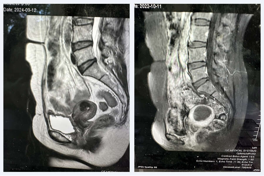
She underwent a uterine artery embolization (UAE) procedure with Dr Lipman and her symptoms went away completely. Shown below are first the sagittal (side view) without contrast and then with intravenous contrast (Figure 2). What is immediately apparent is the significant reduction in the size of the uterus and the area of adenomyosis (Figure 2, left-sided image).
In addition, after intravenous contrast is given, the uterus is alive and enhances brightly with the contrast (i.e. rich blood supply), whereas the small infarcted area of previous adenomyosis does not enhance, i.e. dead, no blood supply (Figure 2 right-hand image).
Uterine Artery Embolization Brings Complete Relief
Not surprisingly, all of the patient’s symptoms had disappeared completely and she was very grateful that she got the relief she was looking for and was able to keep her uterus!
Her Story Doesn’t End There: A Reassuring Follow-Up
Two and a half years later, the patient returned to see Dr. Lipman. She had recently seen her Gynecologist who obtained an ultrasound in his office to “check on the fibroid”. He told the patient that the fibroid was still there and that she still needed a hysterectomy.
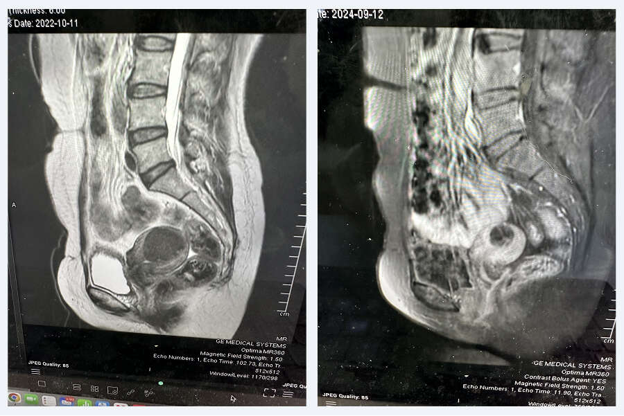
She was confused and reached out for an additional appointment with Dr. Lipman. She asked him if she now needed a hysterectomy or if could she get the embolization again. The first question Dr. Lipman asked was “Have your symptoms returned?” The patient said, “No, not at all. I’m doing great!” “Then you don’t need to be embolized again and you don’t need a hysterectomy either”, he replied. Shown below are the MRI images just prior to that visit showing the return of a normal-sized uterus, along with an ever-shrinking, tiny residual cavity where the adenomyosis used to be (Figure 3).
Seek a Second Opinion with the Atlanta Fibroid Center
If you’re facing a similar diagnosis or being advised to undergo a hysterectomy, it’s essential to explore all your options. Dr. John Lipman and the team at the Atlanta Fibroid Center specialize in nonsurgical treatments like Uterine Artery Embolization (UAE) and can provide a thorough evaluation of your condition. Don’t hesitate to seek a second opinion and take control of your health journey. Contact the Atlanta Fibroid Center today to schedule your consultation.


